
DIAGNOSTIC IMAGING REFERRALS
Detailed assessment for precise diagnosis
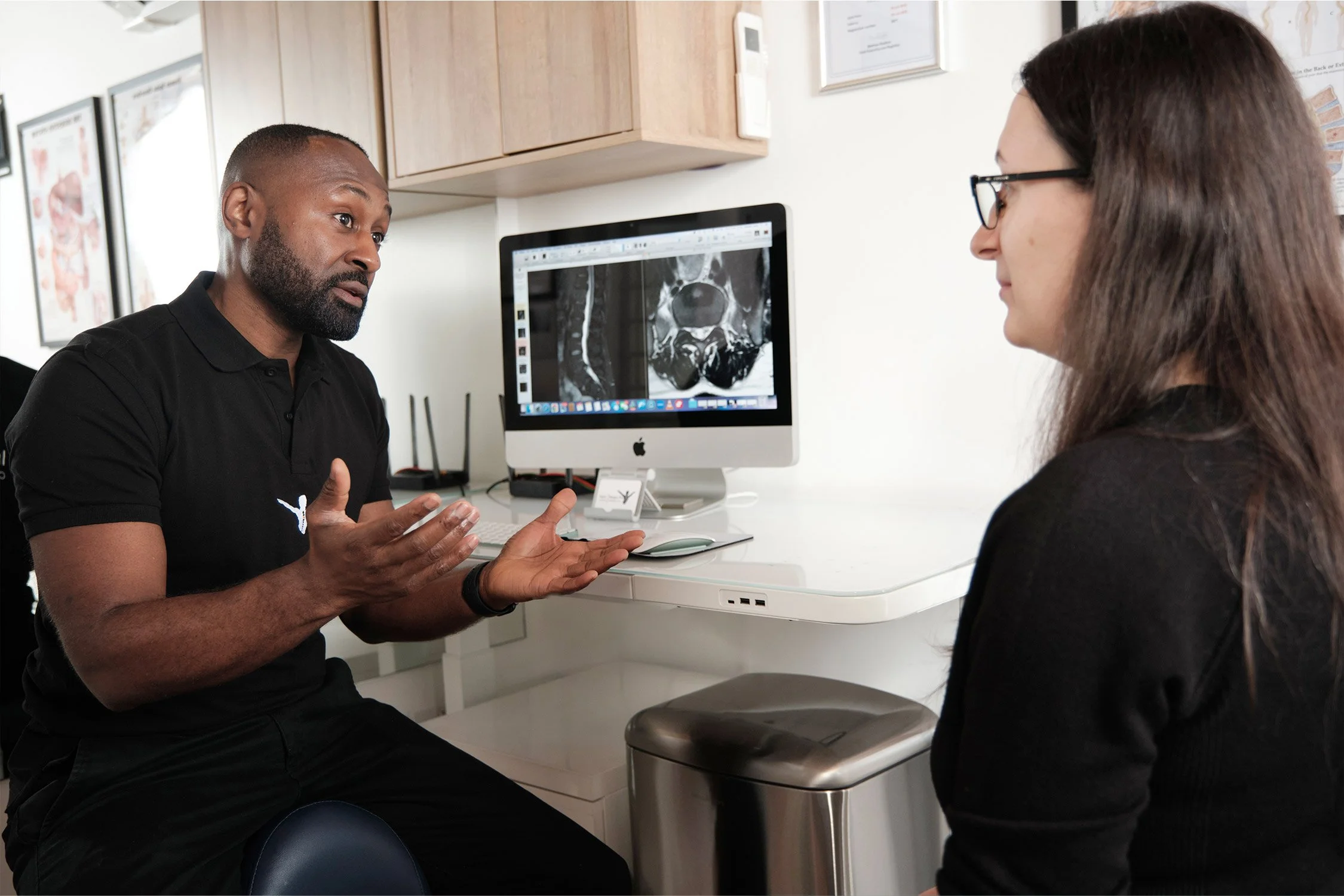
Diagnostic imaging is where specific scans of the body are taken to help create a clearer picture of a case or situation. Images by themselves can be misleading or ambiguous, but when used to inform accurate clinical assessments, they can often help to diagnose and clarify more tricky cases. This means that a specific treatment plan can be put in place to resolve pain, injury and dysfunction more quickly.
We are able to initiate fast referrals for private medical imaging, where we can then go through the results with you and determine the best course of progressive action to take. This method has lead us to the diagnosis of multiple conditions that have previously been missed by other less integrated approaches.
Below are brief explanations of some types of imaging we often refer patients for. We will always discuss with you what we believe is the most suitable scan for your case.
MRI
Magnetic Resonance Imaging. One of the most advanced imaging techniques available, these scans use powerful magnets to create image slices of inside the body in different planes. They are suitable for every body part including bones, soft tissues and the brain, and there is no radiation involved. Scans are performed by laying on a table which goes into a large tube. Seated and open scanners are available for those less mobile or claustrophobic.

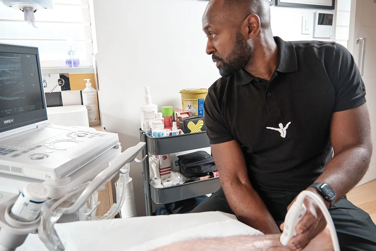
ULTRASOUND
An ultrasound is a scan that uses a probe over the surface of an area to emit high frequency sound waves into the body. These waves bounce off the internal structures scanned to form an image viewable on a monitor. It is non-invasive and very effective at viewing muscles/tendons, thyroid, abdominal contents, pelvic contents, prostate and testicular structures, kidneys and urinary structures.
X-RAY
X-Rays are quick and easy scans that are good for taking 2D images of bones, blood vessels, the heart and lungs. These scans expose the patient to small doses of radiation, but are useful in identifying the presence of fractures, infections, and signs of disease in soft tissues and organs.

CT
A Computerised Tomography (CT) is a non-invasive scan that uses specialised x-ray equipment and is performed inside a large scanner. Sometimes a dye is injected or a drink taken before the scan to produce clearer images. It is effective for viewing inflammation, diseases and cancers in blood vessels, bones, organs, soft tissues and muscles.

DEXA
DEXA stands for Dual Energy X-ray Absorptiometry. These scans are good for measuring bone mineral density, for the identification of conditions such as osteopenia, osteoporosis and osteomalacia. It is a non-invasive, painless procedure which uses low dose x-rays. DEXA also measures body fat (including visceral fat - the dangerous kind), lean muscle mass and how they are distributed around the body. This can be a helpful indicator of health and disease.

Ready To Start Your Recovery?
Book your FREE 15min pre-treatment consultation and we will assess what is causing the pain. We’ll then…
✔ Build a Plan to Restore Movement.
✔ Relieve Pain & Improve Your Mobility
✔ Hands-On Assessment & Treatment
Booking takes less than 2minutes — Choose a time that suits you.
Schedule your FREE Initial Consultation today.
Let’s Get Started